LIVE BLOOD MICROSCOPY– WHAT IS IT ALL ABOUT?
Live blood microscopy, is revolutionising many natural health practices all around the world by giving practitioners the edge they need to achieve better results in their practice.
Live blood microscopy is also known as naturopathic microscopy, live blood cell analysis, nutritional microscopy and The Oxidative Stress test.
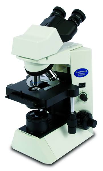
Live blood cell microscopy is the observation of live blood cells using a high powered specialized microscope with a camera that projects a picture of the live blood cells onto a screen to be seen by the practitioner and client together, allowing pictures and videos to be recorded.
A tiny pinprick of blood is put on to a glass slide and then viewed on the screen. In live blood analysis, the blood is not dried or stained beforehand, so the blood elements can be seen in their living state.
The practitioner and client look at the variations in the size, shape, ratio, and fine structure of the red blood cells, white blood cells, platelets, and other blood structures.
The insights gained from the live blood microscopy, correlated with other clinical data, enable the analyst to understand his/her clients individual state of health on a much deeper level.
As a result, an appropriate course of natural treatment and lifestyle/dietary interventions can be formulated and furthermore, the effectiveness of various treatment combinations can be tested and progress can be monitored by observation of changes through further live blood microscopy .
Nutritional microscopy uses live blood analysis with a slant on using nutrition to achieve optimum health naturally.
Dry blood analysis or The Oxidative Stress Test is another very valuable test in live blood cell analysis. With The Oxidative Stress Test (Dry Blood Analysis) a lot can be learned about the level of free radical damage/oxidative stress and toxins in the body.


